
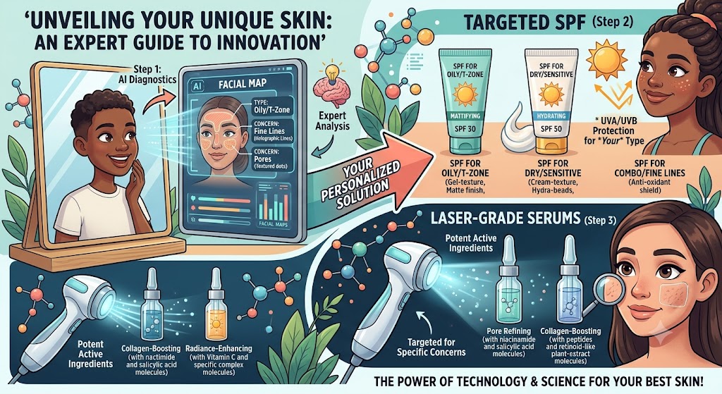
Skin Types & Concerns: Expert Guide to AI Diagnostics, Targeted SPF, and Laser-Grade Serums
The cosmetic dermatology landscape has shifted irrevocably from reactive treatment to preemptive, data-driven architecture. For decades, the beauty industry relied on the four rudimentary skin categories established by Helena Rubinstein in the early 1900s: dry, oily, combination, and sensitive. This legacy model is not just dated; it is scientifically deficient. Human skin operates as a complex, living neuro-immuno-cutaneous organ governed by genetics, environmental epigenetics, hormonal fluxes, and fluctuating barrier dynamics.
Today, the intersection of clinical artificial intelligence (AI), advanced photobiology, and biomimetic peptide chemistry allows us to diagnose and address skin pathology with sub-surface precision. Navigating this new frontier requires looking beyond marketing hyperbole to understand how deep-learning diagnostic algorithms process chromatic variance, why modern sunscreens function as multi-pathway shields rather than passive chalky films, and what truly elevates a topical formulation to “laser-grade” performance.
—
Beyond the Counter: The Modern Taxonomy of Skin Assessment
To implement targeted interventions, we must first abandon generic categorizations. Modern clinical practice leans heavily on the Baumann Skin Typing System, which evaluates skin across four distinct, independent parameters:
- Hydration: Oily (O) vs. Dry (D) — determined by sebum production and sebaceous gland density, not superficial water content.
- Sensitivity: Sensitive (S) vs. Resistant (R) — characterized by the presence or absence of inflammation, barrier impairment, vascular reactivity, and neurosensory hyper-responsiveness.
- Pigmentation: Pigmented (P) vs. Non-Pigmented (N) — measuring the propensity for hyperpigmentation, melanocytic hyperactivity, and post-inflammatory erythema (PIE) via the tyrosinase cascade.
- Elasticity/Wrinkle-Proneness: Wrinkle-Prone (W) vs. Tight (T) — quantifying intrinsic versus extrinsic aging markers, matrix metalloproteinase (MMP) expression, and structural collagen loss.
When evaluated across these axes, patients fall into one of 16 distinct phenotypes. A patient with OSPW (Oily, Sensitive, Pigmented, Wrinkle-Prone) skin requires an entirely inverse molecular strategy compared to one with DRNT (Dry, Resistant, Non-Pigmented, Tight) skin. Applying a rich lipid cream loaded with occlusives to an OSPW barrier will exacerbate follicular occlusion and inflammatory signaling, whereas treating DRNT skin with aggressive keratolytics will swiftly induce secondary transepidermal water loss (TEWL) and barrier collapse.
Overlaying this classification is the Fitzpatrick Phototyping Scale (Types I through VI), which dictates cutaneous response to ultraviolet radiation and heat. In Fitzpatrick Types IV through VI, the melanocytes contain larger, more dispersed melanosomes that react aggressively to mechanical trauma, heat, and UV exposure. Formulating protocols for these skin types demands extreme restraint with thermal devices and low-pH chemical exfoliants to avert refractory Post-Inflammatory Hyperpigmentation (PIH).
—
The AI Diagnostic Revolution: Algorithmic Skin Analysis
Until recently, objective skin assessment was limited to multi-spectral imaging booths like the VISIA system found exclusively in specialty clinical practices. Today, convolutional neural networks (CNNs) and computer vision engines have democratized clinical-grade diagnostics, bringing algorithmic precision directly to smartphones and point-of-care devices.
How Cutaneous AI Diagnostics Operate
Advanced diagnostic platforms rely on deep-learning models trained on millions of clinically annotated dermatological images. These tools decouple superficial skin presentation from underlying pathological activity through three primary mechanisms:
- Cross-Polarized and Parallel-Polarized Simulation: Algorithms can computationally filter out surface glare (specular reflection) to examine sub-surface vasculature, erythema, and telangiectasia. Conversely, parallel-polarization modeling maps surface texture, pore dilation, and micro-relief irregularities.
- Melanin and Hemoglobin Indexing: By analyzing the absorption and scattering of light across red, green, and blue (RGB) color channels, the AI separates melanin deposits (lentigines, melasma, PIH) from oxyhemoglobin pooling (rosacea, persistent erythema, active acne lesions).
- Porphyrin and Bacterial Mapping: Metabolic byproducts of Cutibacterium acnes, known as coproporphyrins, fluoresce under specific spectral bands. AI models can extrapolate bacterial colonization levels within pilosebaceous units by identifying these optical markers before visible comedones or papules emerge.
| Diagnostic Parameter | Traditional Subjective Method | AI-Powered Computer Vision | Clinical Significance |
|---|---|---|---|
| Barrier Integrity (TEWL) | Visual dryness, patient-reported tight sensation | Micro-fissure mapping and corneocyte desquamation indexing | Directs the ratio of ceramides, cholesterol, and free fatty acids needed for repair. |
| Sub-Surface Pigment | Visible spots under daylight inspection | UV-spectrum pigment pooling and dermal nest identification | Preempts visible melasma flares; dictates whether to deploy direct or indirect tyrosinase inhibitors. |
| Vascular Reactivity | Flushing identification post-trigger | Erythema progression heatmaps and capillary dilation indices | Differentiates true barrier degradation from neurogenic rosacea. |
| Biological Age vs. Chronological | Static wrinkle counting | Structural anisotropy, dermal vectoring, and elastosis grading | Benchmarks efficacy of retinoids, growth factors, and collagen-stimulating modalities. |
Clinical Realism: What AI Can and Cannot Do
While AI diagnostic tools excel at tracking treatment progression, optimizing at-home formulations, and identifying sub-clinical solar damage, they are not diagnostic dermatologists. An algorithm can quantify the surface area of hyperpigmentation or count inflammatory papules, but it cannot definitively diagnose amelanotic melanoma, perform a histopathological biopsy of an atypical lesion, or evaluate systemic autoimmune manifestations like lupus erythematosus.
Clinical Boundary: Treat consumer-facing and clinic-grade diagnostic apps as precision monitoring tools. Any evolving, asymmetrical, irregularly bordered, or multicolored lesion must be evaluated in person by a board-certified dermatologist.
—
Targeted Photoprotection: Next-Generation SPF Architecture
The standard recommendation to “wear broad-spectrum SPF 30” is fundamentally incomplete. Modern photobiology demonstrates that solar-induced cutaneous damage extends far beyond the sunburn-inducing UVB spectrum (290–320 nm) and structural UVA-II/UVA-I rays (320–400 nm). High Energy Visible light (HEV/Blue Light, 400–500 nm) and Infrared-A (IRA, 760–1440 nm) penetrate deeply into the reticular dermis, generating reactive oxygen species (ROS), triggering matrix metalloproteinases, and inducing persistent, recalcitrant pigmentation, particularly in Fitzpatrick IV–VI skins.
The Spectrum Breakdown
- UVB (290–320 nm): Absorbed primarily in the epidermis. Causes direct DNA cross-linking (pyrimidine dimers) and acute erythema.
- UVA-I & UVA-II (320–400 nm): Penetrates deep into the dermis. Drives indirect oxidative stress, degrades elastic fibers, suppresses cutaneous immune response, and activates dermal melanocytes.
- HEV / Visible Blue Light (400–500 nm): Generates profound oxidative damage comparable to UVA. In darker phototypes, HEV stimulates a distinct, long-lasting hyperpigmentation complex via the OPN3 (Opsin-3) photoreceptor pathway that traditional chemical and non-tinted mineral filters cannot block.
- Infrared-A (760–1440 nm): Penetrates the subcutis. Up-regulates MMP-1 and MMP-12, directly degrading structural collagen while generating thermal stress within microvascular structures.
Formulation Chemistry: Mineral, Organic, and Hybrid Deployments
The historical debate over mineral versus chemical filters has been surpassed by advances in organic chemistry and particulate engineering. Zinc oxide ($\text{ZnO}$) and Titanium dioxide ($\text{TiO}_2$) remain gold standards for reactive, post-procedural, or rosacea-prone barriers because they are inert, photostable, and non-sensitizing. However, un-tinted micronized mineral filters do not provide protection past 400 nm.
To combat the full spectrum, modern targeted SPFs integrate the following components:
- Iron Oxides (Yellow, Red, Black): Non-negotiable for melasma and hyperpigmentation-prone phenotypes. When combined with zinc oxide, iron oxides extend attenuation into the 400–500 nm HEV spectrum, halting Opsin-3 mediated melanogenesis.
- Modern Chemical Filters (Ex-US / Novel Formulations): Filters such as Bis-Ethylhexyloxyphenol Methoxyphenyl Triazine (Tinosorb S), Methylene Bis-Benzotriazolyl Tetramethylbutylphenol (Tinosorb M), and Diethylamino Hydroxybenzoyl Hexyl Benzoate (Uvinul A Plus) provide photostable, broad-spectrum coverage with high molecular weights, mitigating the irritation potential common with older filters like oxybenzone and avobenzone.
- Endogenous Antioxidant Networks: Modern SPFs are bio-active vehicles. Formulations now integrate lipophilic and hydrophilic free radical quenchers, such as Ectoin (an extremolyte that protects cellular membranes against thermal and osmotic stress), L-Ergothioneine, and Astaxanthin, to neutralize the ROS cascade initiated by breakthrough Infrared-A and visible light.
Choosing SPF by Clinical Indication
- Post-Laser / Compromised Barrier: 100% non-nano zinc oxide (minimum 15-20%) suspended in an anhydrous or high-lipid emulsion containing synthetic ceramides and bisabolol. Zero fragrance, zero volatile silicones, zero chemical filters.
- Recalcitrant Melasma & PIH: Hybrid or high-mineral SPF containing a minimum of 3.5% iron oxides to ensure visible-light block, paired with encapsulated tranexamic acid or niacinamide to downregulate the melanosome transfer process while blocking UV triggers.
- Acne-Prone & Rosacea Subtype 1 (Vascular): Ultralight fluids utilizing modern organic filters or surface-treated zinc oxide, combined with azelaic acid or potassium azeloyl diglycinate. Avoid heavy concentrations of butyloctyl salicylate (a common chemical booster used in “pure mineral” SPFs that can generate heat in vascularly reactive skin).
—
Deciphering “Laser-Grade” Serums: Molecular Realities
The term “laser-grade” is widely used across clinical skincare marketing, but it carries no formal regulatory definition. In clinical practice, a serum earns this designation only if it meets three strict criteria:
- It possesses an advanced transdermal delivery mechanism that bypasses the hydrophobic stratum corneum without disrupting structural lipids.
- It contains clinically proven, bio-identical signaling molecules at active, verified concentrations.
- It demonstrates measurable histopathological alterations in dermal remodeling, mimicking or enhancing the structural outcomes of energy-based devices (e.g., fractional non-ablative lasers, picosecond platforms, or microfocused ultrasound).
Active Profiles of Clinical-Grade Topicals
When engineering a targeted regimen, look for ingredients with verified clinical mechanisms over vague marketing claims:
1. Bio-Identical Peptides and Neuropeptides
Signal peptides like Palmitoyl Tripeptide-1 and Palmitoyl Tetrapeptide-7 (Matrixyl 3000) bind to specific cell surface receptors, triggering the de novo synthesis of Collagen Types I, III, and IV, as well as hyaluronic acid. Copper Tripeptide-1 (GHK-Cu) acts as a powerful remodeling signal, stimulating metalloproteinases to clear damaged, cross-linked collagen bundles while simultaneously stimulating healthy extracellular matrix production. Neuropeptides like Acetyl Hexapeptide-8 target the SNARE protein complex, offering mild, superficial neuromuscular relaxation that complements in-clinic botulinum toxin treatments.
2. Polynucleotides (PDRN) and Exosomes
The cutting edge of regenerative dermatology involves cell-signaling molecules. Polydeoxyribonucleotide (PDRN)—derived from salmon DNA—binds to the Adenosine $A_{2A}$ receptor, systematically down-regulating pro-inflammatory cytokines ($\text{TNF}-\alpha, \text{IL}-6$), stimulating vascular endothelial growth factor (VEGF) for microvascular tissue perfusion, and accelerating re-epithelialization. Exosomes, meanwhile, act as paracrine messengers, delivering microRNA, messenger RNA, and transcription factors directly into senescent fibroblasts to reprogram cellular aging mechanisms.
3. Encapsulated Retinoids and Dermal Delivery Vectors
Traditional retinoic acid causes dramatic epidermal disruption, shedding, and barrier compromise. Laser-grade formulas utilize advanced lipid nanoparticles, liposomal encapsulation, or next-generation esters (such as Hydroxypinacolone Retinoate) paired with transdermal penetration enhancers. This allows the molecule to bypass the upper stratum corneum and reach dermal retinoid receptors without triggering the inflammatory surface cascades typical of unencapsulated tretinoin.
4. Targeted Depigmenting Agents (Non-Hydroquinone)
To safely disrupt melanogenesis without the risk of exogenous ochronosis, high-performance serums deploy a multi-target approach:
- Tranexamic Acid (2–5%): Inhibits the plasmin/plasminogen pathway, effectively severing the inflammatory communication link between keratinocytes, mast cells, and melanocytes.
- Cysteamine Hydrochloride: A potent aminothiol that inhibits both tyrosinase and peroxidase, while significantly increasing intracellular glutathione levels.
- 4-Butylresorcinol: Exhibits an exceptionally high binding affinity for human tyrosinase, outperforming traditional kojic acid and arbutin at lower concentrations with less skin irritation.
—
Protocol Blueprints for Complex Skin Concerns
To illustrate how AI diagnosis, advanced photoprotection, and high-potency serums integrate into a cohesive strategy, consider the following evidence-based protocols designed for three common clinical challenges.
Protocol A: Persistent Vascular Reactivity & Barrier Compromise
Target Profile: Baumann S/R parameter: Highly Sensitive (S). Presents with diffuse facial erythema, elevated TEWL, stinging sensations, and visible capillary ectasia. Often misidentified as simple “dry skin.”
- Morning:
- Cleanse: Non-surfactant lipid emulsion cleanser (containing ceramides and thermal water). Pat dry; do not rub.
- Intervention Serum: High-concentration Centella Asiatica titrated extract (Madecassoside, Asiaticoside) combined with Copper Tripeptide-1 (GHK-Cu) to stimulate microvascular integrity and down-regulate $\text{IL}-8$.
- Moisturizer: Physiological lipid replenisher containing a balanced 3:1:1:1 ratio of ceramides, cholesterol, free fatty acids, and squalane.
- Photoprotection: 100% un-tinted micronized Zinc Oxide ($>18\%$) fortified with Ectoin. Free of drying alcohols, botanical essential oils, and volatile silicones.
- Evening:
- First Cleanse: High-purity squalane or caprylic/capric triglyceride cleansing oil to solubilize mineral SPF without disrupting the acid mantle.
- Second Cleanse: Mild amino acid surfactant gel wash (pH 5.2–5.5).
- Treatment: Prescription-strength Azelaic Acid (15% gel or 20% cream). Inhibits kallikrein-5 and cathelicidin transcription to directly clear inflammatory erythema.
- Repair: Biomimetic barrier balm fortified with colloidal oat extract, panthenol (5%), and synthetic pseudoceramides.
Protocol B: Recalcitrant Melasma & Post-Inflammatory Hyperpigmentation
Target Profile: Baumann Pigmented (P), Fitzpatrick Types III–V. Chronic, deep dermal/epidermal pigment patches triggered by hormone fluctuations, heat, and UV/HEV light.
- Morning:
- Cleanse: Low-pH, non-stripping cleanser.
- Laser-Grade Depigmenting Serum: Multi-pathway formula containing Tranexamic Acid (3%), N-Acetyl Glucosamine (2%), Alpha Arbutin (2%), and 4-Butylresorcinol.
- Antioxidant Shield: Oil-soluble Vitamin C (Tetrahexyldecyl Ascorbate, 10–15%) paired with Ferulic Acid. Avoid low-pH L-Ascorbic acid serums if the barrier shows baseline sensitivity, as the resulting inflammation can provoke secondary melanogenesis.
- Photoprotection: Hybrid or high-mineral broad-spectrum SPF 50+ containing a minimum of 4% Iron Oxides to block High-Energy Visible (HEV) blue light. Reapplication every 2 hours using a tinted mineral powder or fluid cushion.
- Evening:
- Cleanse: Gentle enzymatic or micellar double cleanse.
- Targeted Renewal: Liposomal Retinaldehyde (0.05% to 0.1%) or Hydroxypinacolone Retinoate (HPR). Retinaldehyde accelerates keratinocyte turnover with direct antibacterial and barrier-sparing benefits compared to classic retinol.
- Targeted Inhibitor Layer: Niacinamide (4-5%, avoiding hyper-concentrated 10-20% variants that induce flushing) combined with licorice root extract (glabridin) to halt melanosome transfer to surrounding cells.
- Barrier Lock: Lightweight, cholesterol-rich ceramide gel-cream.
Protocol C: Accelerated Extrinsic Photoaging with Structural Laxity
Target Profile: Baumann Wrinkle-Prone (W). Exhibits dermal elastosis, reduced dermal thickness, fine and dynamic expression lines, and localized collagen depletion.
- Morning:
- Cleanse: Gentle hydrating gel wash.
- Signal Serum: Recombinant Human Growth Factor complex (EGF, FGF, VEGF) combined with Acetyl Hexapeptide-8 and Palmitoyl Tripeptide-38.
- Hydration Network: Multi-molecular weight Hyaluronic Acid paired with Polyglutamic Acid to bind moisture simultaneously at superficial and deep dermal levels.
- Photoprotection: Broad-spectrum SPF 50+ rich in lipophilic antioxidants (Astaxanthin, Coenzyme Q10) to intercept Infrared-A and UVA-driven Matrix Metalloproteinase (MMP) induction.
- Evening:
- Double Cleanse: Conditioning lipid balm followed by a gentle surfactant cleanser.
- Structural Correction: Encapsulated Tretinoin (0.025–0.05%) or Micro-encapsulated Pure Retinol (0.5–1.0%). Start on alternate evenings, buffering with hydration to prevent cellular inflammation.
- Regenerative Recovery: Exosome or PDRN-infused repair serum applied 15 minutes post-retinoid to stimulate DNA repair cascades and optimize dermal remodeling.
- Moisturizer: Biomimetic peptide-infused emollient cream containing palmitoyl-derived matrix-repair complexes.
—
Clinical Boundaries: Cosmeceuticals vs. In-Office Interventions
Skincare protocols, even those featuring sophisticated, laser-grade serums, are fundamentally limited by physical laws. The stratum corneum exists specifically to prevent external macromolecules from penetrating the body. Molecules with a molecular weight greater than 500 Daltons cannot passively diffuse across an intact stratum corneum—a principle known in transdermal pharmacology as the 500 Dalton Rule.
High-molecular-weight hyaluronic acid, native collagen molecules, and whole growth factor proteins cannot penetrate deep into the reticular dermis via simple topical application. They work by hydrating the surface, protecting the upper barrier, and initiating cell-signaling cascades through outer keratinocyte receptors.
True structural skin remodeling requires an integrated, staged strategy:
- Topicals Create the Foundation: Pre-procedural conditioning using targeted SPF, barrier-support lipids, and tyrosinase inhibitors optimizes the skin’s biological resilience. This significantly lowers the risk of post-inflammatory hyperpigmentation (PIH) and speeds up healing times after in-office treatments.
- In-Office Procedures Drive Dermal Architecture: Non-ablative fractional lasers (1550 nm/1927 nm), ablative $\text{CO}_2$ resurfacing, microfocused ultrasound (MFU-V), radiofrequency microneedling, and deep chemical peels bypass the physical 500 Dalton barrier. They deliberately induce controlled micro-thermal injuries that trigger neocollagenesis deep within the dermis—a biological process that no topical serum can match on its own.
- Post-Procedure Topicals Direct the Healing Cascade: Once the stratum corneum is intentionally perforated by microneedles or fractionated thermal zones, the window opens for true deep transdermal delivery. Applying sterile PDRN, recombinant growth factors, and pure biomimetic peptides during this transient window maximizes their regenerative impact.
—
Actionable Implementation Guide: 4-Step Regimen Audit
Before introducing high-performance, laser-grade actives into your routine, optimize your daily regimen by following this structured process:
- Establish Your True Baseline: Remove all active ingredients (exfoliating acids, retinoids, high-percentage L-ascorbic acid) for 72 hours. Assess your skin’s innate sebum production, tightness, and erythema using natural daylight or a validated clinical AI scanning platform.
- Repair and Stabilize the Barrier First: If your skin stings upon applying a neutral moisturizer, your barrier is compromised. Do not apply laser-grade depigmenting serums or encapsulated retinoids to an inflamed baseline. Dedicate two to four weeks entirely to barrier restoration using 3:1:1:1 physiological lipids, zinc oxide sunscreen, and gentle hydrators.
- Integrate Actives Singularly: Introduce high-potency serums one at a time, spaced at least 14 days apart. This deliberate pacing helps you identify the specific cause of any irritant contact dermatitis, breakouts, or sensitivity flares.
- Upgrade Your Sunscreen to Match Your Concern: If hyperpigmentation is your primary issue, immediately switch to an iron-oxide-fortified tinted sunscreen to block blue light. If you struggle with facial redness or rosacea, eliminate volatile organic UV filters and transition to a soothing, pure zinc oxide formula.
—
Frequently Asked Questions
Can AI skin diagnostics accurately detect skin cancer or melanomas?
No. Consumer AI diagnostic applications and computerized beauty scanners are designed solely to evaluate cosmetic metrics: skin hydration, pore surface area, wrinkles, erythema, and superficial pigmentation. They are neither cleared nor calibrated to diagnose cutaneous malignancies. Any changing, asymmetrical, dark, or bleeding lesion must be examined in person by a board-certified dermatologist using high-resolution dermoscopy and histopathological analysis.
What does “laser-grade” actually mean on an ingredient label?
“Laser-grade” is an industry marketing term rather than a formal regulatory standard. It typically describes serums that feature advanced transdermal delivery systems (such as liposomal encapsulation), pharmaceutical-grade purity levels, and clinical testing that mirrors the cellular repair mechanisms targeted by in-office lasers (such as stimulation of collagen, inhibition of melanogenesis, or reduction of inflammation). Always evaluate the formula by its active ingredients and delivery mechanisms rather than front-label claims.
Do I need an iron-oxide sunscreen if I work indoors all day?
If you struggle with melasma, deep post-inflammatory hyperpigmentation, or severe photoaging, yes. Standard window glass blocks UVB and some UVA rays, but it does not block High Energy Visible (HEV) blue light emitted by the sun or interior lighting. Furthermore, device screens emit blue light directly onto facial skin at close range. Iron oxides (red, yellow, and black) are essential because they physically reflect and absorb these visible wavelengths, protecting hyper-reactive melanocytes from unwanted stimulation.
Can growth factors and exosomes in serums replace in-clinic procedures?
No. Topically applied growth factors and exosomes cannot match the dermal structural remodeling achieved by energy-based devices like fractional lasers or radiofrequency microneedling. These clinical platforms bypass the physical stratum corneum barrier to stimulate deep tissue repair. Rather than replacing clinical treatments, topical growth factor serums complement them: they prepare the skin prior to procedures, dramatically accelerate post-treatment re-epithelialization, and help maintain the clinical results over the long term.
How long does it take to see visible results from targeted depigmenting serums?
Melanogenesis is a gradual cellular process. Keratinocytes take approximately 28 to 40 days to migrate from the basal layer to the surface of the stratum corneum, a cycle that lengthens as we age. Because non-hydroquinone depigmenting actives (such as tranexamic acid, cysteamine, and 4-butylresorcinol) work by gently inhibiting ongoing pigment synthesis and transfer rather than aggressively destroying cells, you should expect to use them consistently for 8 to 12 weeks before seeing significant clinical clearance of stubborn pigmentation.
—
Medical Disclaimer: This article is intended solely for educational, editorial, and informational purposes. It does not constitute medical advice, clinical diagnosis, or individualized treatment plans. Always consult a board-certified dermatologist or licensed medical professional before beginning any high-potency cosmeceutical protocol, especially if you have an underlying dermatological condition, active infection, or unresolved skin lesion.

One thought on “Skin Types & Concerns: Expert Guide to AI Diagnostics, Targeted SPF, and Laser-Grade Serums”
Comments are closed.