Ingredient Deep-Dives
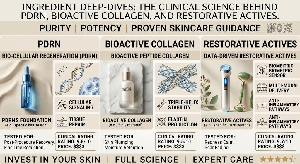
Ingredient Deep-Dives: The Clinical Science Behind PDRN, Bioactive Collagen, and Restorative Actives
For decades, corrective dermatology and cosmetic chemistry operated under a paradigm of controlled micro-trauma followed by passive barrier protection. We ablated the epidermis with high-fluence lasers, induced micro-wounds via mechanical needles, or stripped the stratum corneum with low-pH chemical peels, relying entirely on the skin’s intrinsic biological reserves to rebuild what we deliberately compromised. The supportive topical regimen was modest: petrolatum-based occlusives, basic humectants, and standard antioxidants.
That paradigm is fundamentally obsolete. Today, cellular senescence, chronological aging, chronic low-grade inflammation (“inflammaging”), and environmental oxidative load often leave the cutaneous matrix functionally depleted. Cells exposed to sustained solar elastosis or biological exhaustion do not possess the energetic or structural capital to mount an ideal repair cascade. The consequence is prolonged erythema, dyspigmentation, delayed re-epithelialization, and fibroblastic insufficiency.
This reality has driven the rise of bioregenerative aesthetics: an advanced discipline focused on supplying targeted physiological signals and biological building blocks directly to cutaneous tissue. Leading this pharmacological shift are three classes of restorative molecules: Polydeoxyribonucleotide (PDRN), Bioactive and Recombinant Collagens, and a synergistic suite of cellular-priming Restorative Actives (including GHK-Cu, Ectoin, and Titrated Triterpenes). Understanding how to evaluate, sequence, and formulate with these agents requires moving past marketing jargon to interrogate their receptor kinetics, signaling pathways, and structural behaviors within the extracellular matrix (ECM).
Polydeoxyribonucleotide (PDRN): The Cellular Salvage Engine
Originally derived from the germ cells of Oncorhynchus mykiss (Salmon trout) or Oncorhynchus keta (Chum salmon), PDRN represents a specialized class of DNA-derived therapeutic polymers. The biological rationale for utilizing salmon DNA centers on its high degree of bio-compatibility with human genome sequences (exceeding 95%) and an extraction process designed to yield low-molecular-weight fractions—typically between 50 and 1,500 kilodaltons (kDa)—devoid of immunogenic proteins, lipids, and active viral components.
Primary Mechanism: Adenosine A2A Receptor Agonism
While often mischaracterized merely as a source of “cellular nutrition,” PDRN’s clinical efficacy stems from a specific pharmacodynamic interaction: the selective, sustained agonism of the Adenosine A2A purinergic receptor. Adenosine receptors ($A_1$, $A_{2A}$, $A_{2B}$, and $A_3$) are G-protein-coupled receptors embedded in mammalian cell membranes that modulate vascular, immunological, and regenerative signaling.
Under tissue stress, ischemia, or mechanical trauma, endogenous adenosine release is transient because adenosine deaminase rapidly degrades it into inosine. PDRN bypasses this limitation. By engaging the $A_{2A}$ receptor over an extended therapeutic window, PDRN initiates a downstream signaling cascade that suppresses pro-inflammatory cytokines while elevating tissue-reparative mediators:
- Suppression of Cytokine Storm Dynamics: Downregulation of Tumor Necrosis Factor-alpha (TNF-α), Interleukin-6 (IL-6), and high-mobility group box 1 (HMGB-1) protein.
- Amplification of Reparative Cytokines: Upregulation of Interleukin-10 (IL-10), the body’s premier anti-inflammatory mediator.
- Vascular Endothelial Growth Factor (VEGF) Induction: Promoting controlled neovascularization and micro-angiogenesis to restore capillary perfusion to ischemic, aged, or wounded dermis.
Secondary Mechanism: The Nucleoside Salvage Pathway
Beyond receptor-mediated signaling, enzymatic cleavage of PDRN yields free deoxyribonucleotides, nucleosides, and nitrogenous bases. Cells possess two paths to synthesize DNA during mitosis and cellular repair: the energy-intensive de novo synthesis pathway and the salvage pathway.
De novo synthesis demands substantial adenosine triphosphate (ATP) expenditure, requiring the progressive construction of purine and pyrimidine rings from amino acids and phosphoribosyl pyrophosphate. In senescent, ischemic, or UV-irradiated fibroblasts, metabolic fatigue slows this process down. By supplying readymade nucleotide precursors, PDRN fuels the salvage pathway, allowing cells to salvage and integrate purines and pyrimidines into newly minted genomic material with a fraction of the metabolic expenditure. This accelerates DNA repair, supports cellular proliferation, and drives physiological re-epithelialization without draining the cell’s energetic reserves.
Clinical Profile: Topical Permeation vs. Injectable Mesotherapy
In aesthetic medicine, PDRN is celebrated globally through intradermal formulations (such as Rejuran). Intradermal delivery deposits the polymer directly into the papillary dermis, where fibroblast density and vascular interaction are highest. This approach provides rapid improvements in dermal thickness, skin elasticity, and microrelief.
Topically, PDRN presents a biophysical challenge: its molecular weight, despite fragmentation, remains substantially higher than the traditional 500-Dalton threshold for passive stratum corneum transit. Left unassisted, topical high-mass PDRN operates primarily as a humectant and immediate post-barrier repair agent. However, true transdermal delivery of PDRN can be achieved through three main strategies:
- Permeation Enhancers & Liposomal Nanocarriers: Encapsulation within elastic, deformable liposomes (transfersomes) or solid lipid nanoparticles that can navigate the tortuous lipid bilayer pathways of the intercorneocyte space.
- Chemical Fragmenting: Specialized low-molecular-weight PDRN fragments (down to 20–50 kDa oligodeoxynucleotides) engineered to slip through transient follicular conduits.
- Fractional Transdermal Delivery: Applying high-purity PDRN immediately following micro-needling, non-ablative fractional laser (NAFL), or radiofrequency micro-needling. Clinical micro-channels entirely bypass the physical resistance of the stratum corneum, allowing topically applied PDRN direct access to the viable epidermis and upper papillary dermis, matching the histological outcomes of classical mesotherapy.
Bioactive Collagen: Transcending the Moisture Barrier Myth
For half a century, the inclusion of “collagen” on an ingredient label was a classic hallmark of skincare marketing hyperbole. Native animal collagen (predominantly derived from bovine hides, porcine dermis, or fish scales) exists as a high-molecular-weight triple helix measuring roughly 300,000 Daltons (300 kDa) with dimensions near 300 nanometers long and 1.5 nanometers wide. Applying a native 300 kDa helical structural protein to human skin yields an exceptional non-occlusive film-forming agent that binds surface water effectively, but it cannot penetrate the stratum corneum to replace missing dermal collagen fibers.
Recent advances in recombinant biotechnology and molecular enzymatic processing have changed this reality. The industry has advanced beyond whole animal extractions to embrace Bioactive Peptides, Hydrolyzed Biological Fractions, and Recombinant Human (rh) Collagen.
Recombinant Human Collagens (rh-Collagen Type I, III, and XVII)
Engineered via synthetic biology through eukaryotic yeast expression systems (such as Pichia pastoris), bacteria (E. coli), or plant vectors (transgenic Nicotiana benthamiana), rh-Collagens represent a paradigm shift in biomimetics. These proteins are not animal derivatives; they are constructed with human-identical genetic sequencing.
Crucially, rh-Collagen can be synthesized as specific, active structural motifs or fragments precisely scaled to target sizes:
- rh-Collagen Type I: Focuses on restoring tensile strength to the mature reticular dermis, anchoring the broad extracellular framework.
- rh-Collagen Type III: Often called “baby collagen” or embryonic collagen. This isoform forms compliant, elastic fibrillar networks alongside Type I. In adult skin, the Type I-to-Type III ratio shifts from a youthfully resilient balance toward a rigid, Type-I dominated matrix. Reintroducing biomimetic Type III signals triggers tissue compliance and mitigates scarring.
- rh-Collagen Type XVII (COL17A1): A transmembrane collagen localized to the dermal-epidermal junction (DEJ), specifically within hemidesmosomes. Type XVII collagen acts as the direct master regulator of hair follicle stem cells and epidermal stem cell competition. Loss of COL17A1 leads to stem cell senescence and subsequent epidermal thinning. Advanced restorative topicals now integrate specific Type XVII-mimetic peptide motifs to anchor basal keratinocytes and stabilize the basement membrane.
Peptide Signaling Motifs: The Direct Cellular Messengers
When collagen undergoes targeted enzymatic hydrolysis, it yields specific bioactive di- and tripeptides that function not as structural fillers, but as primary matrikines—signaling fragments liberated during matrix breakdown that command cells to orchestrate renewal. The most biologically potent of these are sequences containing proline and hydroxyproline, specifically:
- Prolyl-hydroxyproline (Pro-Hyp): A dipeptide that binds directly to cell-surface receptors on dermal fibroblasts, stimulating cell migration and triggering the endogenous synthesis of hyaluronic acid via Hyaluronan Synthase-2 (HAS2) upregulation.
- Glycyl-prolyl-hydroxyproline (Gly-Pro-Hyp): A tripeptide that serves as an unambiguous biomarker of matrix turnover, driving the transcriptional upregulation of procollagen Type I C-peptide (PIP).
These peptides engage with integrin receptors (specifically the $\alpha_1\beta_1$ and $\alpha_2\beta_1$ heterodimers) located on the outer fibroblast membrane. This binding event initiates an intracellular signaling cascade: phosphorylation of focal adhesion kinase (FAK), followed by the activation of the Mitogen-Activated Protein Kinase (MAPK) and Extracellular Signal-Regulated Kinase (ERK) cascades. The ultimate cellular outcome is clear: the fibroblast interprets the presence of these precise fragments as an active ECM crisis, responding with fresh, endogenous collagen and glycosaminoglycan synthesis.
The Restorative Actives Symphony
PDRN and bioactive collagens rely on an optimal cellular microenvironment to function effectively. If a patient’s tissue microenvironment is compromised by oxidative stress, hyperosmotic shock, or hyperactive matrix metalloproteinases (MMPs), these signaling agents face immediate degradation. Sustained cutaneous restoration demands pairing them with targeted, biologically potent co-actives.
1. GHK-Cu (Copper Tripeptide-1)
Isolated originally from human plasma, GHK (glycyl-L-histidyl-L-lysine) is a naturally occurring peptide sequence with a remarkably high binding affinity for Copper (II) ions ($Cu^{2+}$). While conventionally labeled a “collagen booster,” GHK-Cu is more accurately characterized as an ECM Remodeling Conductor.
Its primary therapeutic genius lies in its bidirectional regulation of tissue architecture. Chronically aged or scarred tissue is filled with chaotic, non-functional, cross-linked structural proteins, alongside excessive localized MMP levels that destroy newly synthesized proteins. GHK-Cu restores equilibrium:
- It upregulates tissue inhibitors of metalloproteinases (TIMP-1 and TIMP-2), suppressing uncontrolled ECM degradation.
- Concurrently, it modulates specific MMPs (MMP-1, MMP-2) to dismantle abnormal scar matrix, clearing space for fresh, ordered fibrillogenesis.
- It demonstrates anti-senescence properties by downregulating markers of cellular arrest and encouraging stem cell proliferation in the basal layer of the epidermis.
2. Ectoin: The Extremolyte Shield
Derived from halophilic extremophilic microorganisms that thrive in salt lakes, hydrothermal vents, and arid deserts, Ectoin is a zwitterionic cyclic amino acid derivative (1,4,5,6-tetrahydro-2-methyl-4-pyrimidinecarboxylic acid). It operates entirely outside traditional receptor pathways, utilizing physical chemistry to preserve biological integrity.
Ectoin is a premier osmolyte. Because of its unique dipolar structure, it binds water molecules tightly, constructing an ultra-dense, stable aqueous envelope around cell membranes, protein complexes, and cellular enzymes—a phenomenon known as the preferential exclusion mechanism. This hydration mantle shields skin cells from:
- Surfactant-induced protein denaturation.
- Thermal stress (heat shock protein activation).
- UVA- and particulate matter-induced cellular membrane oxidation.
- Transepidermal water loss (TEWL) driven by osmotic collapse during acute barrier compromise.
By protecting structural proteins against dehydration-induced misfolding, Ectoin guarantees that freshly supplied bioactive collagens and cytokines retain their tertiary, biologically active shape upon dermal application.
3. Titrated Extract of Centella Asiatica (TECA)
Standard “Centella extract” is often little more than a dilute water-and-glycerin solution with minimal bioactive retention. By contrast, the gold standard for clinical repair is TECA: a highly purified, pharmaceutical-grade mixture of three distinct triterpenic molecules: Asiaticoside (approx. 40%), Madecassic Acid (approx. 30%), and Asiatic Acid (approx. 30%).
The molecular mechanics of these three triterpenes function with notable synergy:
- Asiaticoside: The master driver of angiogenesis and collagen synthesis. It actively binds the TGF-$\beta$ receptor type I (T$\beta$R-I), inducing Smad2/3 phosphorylation and subsequently driving high-efficiency transcription of Type I collagen.
- Madecassic and Asiatic Acids: Specialized anti-inflammatory agents that suppress the NF-$\kappa$B transcriptional pathway. They directly downregulate inducible Nitric Oxide Synthase (iNOS) and Cyclooxygenase-2 (COX-2), extinguishing chronic, low-grade sub-clinical inflammation that otherwise degrades the newly generated collagen matrix.
4. Epidermal Growth Factor (rh-Oligopeptide-1)
Epidermal Growth Factor (EGF) is a 53-amino-acid polypeptide that binds directly to the EGFR (ErbB-1) on the membrane of basal keratinocytes and fibroblasts. Once bound, it activates intrinsic receptor tyrosine kinase activity, sparking the cell division, migration, and survival programs required for rapid re-epithelialization following physical trauma.
Clinical caution must be exercised with Growth Factors. Unchecked mitogenic stimulation is undesirable in tissue showing premalignant signs (such as severe actinic keratoses). However, within acute, controlled wound environments or severely atrophic skin, high-purity recombinant human EGF acts as an effective cellular accelerator, signaling keratinocytes to rapidly close surface gaps while PDRN and Bioactive Collagen rebuild the underlying dermis.
Comparative Mechanics: Bioregenerative Actives Matrix
To construct rational, evidence-based topical formulations or clinical procedures, clinicians and cosmetic chemists must look at the distinct molecular weight, primary receptor targets, and biological endpoints of each restorative agent:
| Active Component | Average Molecular Weight | Primary Target / Receptor | Key Biological Endpoint |
|---|---|---|---|
| PDRN | 50 – 1,500 kDa | Adenosine A2A Receptor; Nucleoside Salvage | Anti-inflammatory (IL-10 ↑), micro-circulation (VEGF ↑), cellular salvage ATP efficiency. |
| rh-Collagen (Type III) | 5 – 100 kDa (Engineered) | Integrins (α1β1, α2β1) | Signals ECM restructuring; improves mechanical elasticity and compliant cellular scaffolding. |
| GHK-Cu | ~340 Da (Free peptide) | Direct genomic transcription; copper transport | Balances MMP degradation with collagen synthesis; downregulates senescent secretory phenotype. |
| Ectoin | ~142 Da | Cellular water dipole matrices; physical exclusion | Membrane stabilization, osmotic normalization, environmental stress and protein fold shielding. |
| TECA | ~450 – 960 Da | TGF-β Receptor Type I; NF-κB pathway | Suppression of pro-inflammatory enzymes (COX-2); upregulates structural procollagen transcription. |
Formulation Dynamics and Delivery Systems: Overcoming the Barrier
The central hurdle of modern restorative skincare is not biological efficacy at the cellular level; it is the physical delivery of these macromolecules past the 15-micrometer hydrophobic brick-and-mortar architecture of the stratum corneum. The 500-Dalton rule remains an undeniable biological reality: molecules with a molecular mass above 500 Da, especially those possessing hydrophilic charges, demonstrate minimal passive diffusion across intact skin.
1. Biomimetic Liposomal & Deformable Vesicle Encapsulation
For non-invasive topical applications, macromolecules like PDRN, rh-Collagen, and hydrophilic peptides must be shielded within advanced carrier systems. Conventional liposomes (rigid phospholipid bilayers) often rupture in the superficial layers of the stratum corneum, releasing their cargo on the surface. To circumvent this, the cosmetic sciences have developed elastic vesicles (transfersomes) and ethosomes.
These specialized carriers incorporate chemical edge-activators (such as polysorbate surfactants or specific ratios of ethanol) that destabilize the vesicle’s lipid membrane just enough to confer immense structural flexibility. When driving through the tortuous interstitial pathways between corneocytes, these vesicles deform under osmotic gradient forces without rupturing, transporting complex biological cargo into the deeper, viable nucleated epidermis and upper dermis.
2. Micro-Channel Optimization (Physical Delivery)
The definitive delivery system for high-molecular-weight restorative actives remains mechanical micro-puncture. When using a professional automated micro-needling device or a fractionated non-ablative laser, thousands of transient aqueous channels are pierced through the hydrophobic stratum corneum directly into the vascularized, cellular papillary dermis.
Applying an unpreserved, sterile solution of PDRN and recombinant collagen during this precise interventional window turns low-bioavailability topicals into functional intra-tissue biological modulators. Within these micro-channels, the molecules encounter fibroblasts and capillary networks directly, driving rapid clinical responses within hours rather than months.
3. Chemical Incompatibilities and Formulation Stability
Formulating with complex restorative actives introduces real stability challenges. Chemists must account for three common incompatibilities:
- Chelator Incompatibility with GHK-Cu: Standard formulations often use Disodium EDTA or Tetrasodium Glutamate Diacetate as heavy metal chelators. However, introducing EDTA to a GHK-Cu formulation strips the $Cu^{2+}$ ion straight out of the peptide complex, rendering the active completely inert and collapsing its characteristic blue tint.
- pH Inversion and Collagen Unfolding: Recombinant human collagens and delicate oligopeptides degrade when exposed to aggressive pH environments. Pairing low-pH chemical exfoliants (such as 15% Glycolic or Salicylic acid at pH 3.2) in the exact same layer as rh-Collagen or PDRN will hydrolyze peptide backbones and denature structural motifs, rendering them biologically unrecognized by cellular integrins. Restorative complexes perform best within a strictly buffered physiological pH bracket between 5.5 and 6.8.
- Enzymatic Breakdown: High biological activity formulations require rigorous preservation systems that do not rely on destabilizing alcohol profiles, which precipitate and deactivate delicate high-molecular-weight proteins.
Translating the Science: Evidence-Based Clinical Application
To apply these clinical insights efficiently, practitioners and clinical skin specialists should avoid stacking unstable biologicals indiscriminately. High-efficacy restoration demands targeted sequencing that respects molecular weights, structural dynamics, and the acute inflammatory phase of healing.
Protocol 1: Post-Ablative or Post-Needling Acute Regeneration
Immediately following in-clinic procedures that deliberately disrupt the epidermal barrier (e.g., fractional laser, RF-needling, medium-depth chemical peels):
- Phase 1: Direct Signaling (Within 0 to 4 hours post-procedure). Apply sterile, preservative-free, non-crosslinked high-viscosity PDRN along with low-molecular-weight rh-Collagen Type III directly into open micro-channels. At this stage, avoid complex emulsions, fragrance, heavy occlusive silicones, or chemical preservatives that can form foreign-body granulomas in the open dermis.
- Phase 2: Osmotic and Inflammatory Calming (Day 1 through Day 3). Introduce a clean aqueous suspension combining Ectoin (1.0–2.0%), TECA (0.5–1.0%), and high-potency non-animal bio-peptides. This combination establishes a dense hydration shell over damaged cellular membranes, suppresses excessive NF-$\kappa$B mediated swelling, and accelerates keratinocyte bridge closure.
- Phase 3: ECM Maturation (Day 4 onward). Once the stratum corneum re-epithelializes, transition to a stabilized delivery cream containing encapsulated GHK-Cu (1.0%) and physiological biomimetic lipids (ceramides, cholesterol, free fatty acids in a 3:1:1 ratio) to clear disorganized matrix debris and establish balanced, functional dermal architecture.
Protocol 2: Managing Chronic Inflammaging and Biological Exhaustion
For mature, thin, UV-damaged skin suffering from reduced dermal thickness and broken, elastotic capillaries:
- Morning Routine (Protection & Signal Amplification): Cleanse with a non-stripping, neutral-pH surfactant. Apply an active serum containing Ectoin (2%) combined with a biomimetic rh-Collagen Type XVII and Type I fragment complex. Top with an antioxidant (such as Ascorbyl Glucoside or Ergothioneine) and finish with a broad-spectrum, high-UVA-PF sunscreen to protect against environmental degradation of newly produced matrix proteins.
- Evening Routine (Metabolic Repair & Remodeling): Alternate application evenings. On Night A, apply an encapsulated PDRN transfersomal serum, allowing the nucleosides to feed the salvage pathway during natural overnight cell division. On Night B, apply a stabilized GHK-Cu and TECA emulsion, keeping retinoids to a targeted, well-tolerated schedule (e.g., 2 nights a week) to prevent overlapping cellular exhaustion or excessive barrier disruption.
Key Takeaways for the Clinician and Formulator
- PDRN is not just an anti-inflammatory humectant: It serves as a biological agonist for the Adenosine A2A receptor that simultaneously upregulates VEGF and IL-10 while fueling the cellular salvage pathway to repair genomic structural damage without high ATP consumption.
- Native animal collagen does not remodel the dermis: True dermal restoration requires advanced recombinant human collagens (rh-Collagen I, III, XVII) and targeted matrikines (Pro-Hyp, Gly-Pro-Hyp) that actively trigger integrin signaling cascades to stimulate natural procollagen transcription.
- Restorative actives demand structural synergy: GHK-Cu regulates MMP-driven structural degradation; Ectoin builds an osmoprotective physical water shell protecting proteins from external stressors; and pharmaceutical-grade TECA downregulates underlying inflammatory cytokines via the TGF-$\beta$ signaling pathway.
- Molecular size dictates formulation design: Without micro-channel delivery (such as micro-needling) or flexible nanocarrier encapsulation (such as transfersomes or ethosomes), large-molecule PDRN and full-chain collagens stay limited to the stratum corneum surface. Always balance formulation pH strictly between 5.5 and 6.8 to prevent peptide and structural denaturation.
Frequently Asked Questions
Can PDRN and Bioactive Collagen be combined with prescription Retinoids?
Yes, and they are functionally complementary when sequenced properly. Retinoids operate primarily by binding to nuclear retinoic acid receptors (RAR/RXR), inducing rapid keratinocyte proliferation and increasing fibroblast activity. However, they frequently cause localized irritation, barrier breakdown, and transepidermal water loss. Introducing PDRN and Ectoin dampens retinoid-induced neuro-inflammation via purinergic signaling, while bioactive collagen fragments provide immediate support for the high structural turnover triggered by retinoid application. For patients with sensitized skin, alternate nights: apply the retinoid on Night 1 and the restorative complex on Night 2.
Is Salmon-derived PDRN safe for individuals with severe seafood allergies?
In clinical literature, high-purity medical-grade PDRN has exhibited virtually zero cross-reactivity with dietary fish or seafood allergies. This safety profile is rooted in the extraction and purification methodology: the manufacturing process extracts, isolates, and purifies ultra-specific, low-molecular-weight DNA fractions while enzymatically degrading and stripping away the proteins (specifically parvalbumin and tropomyosin) and cellular lipids that trigger type-I IgE-mediated allergic hypersensitivity reactions. Nevertheless, for patients with a documented history of severe anaphylaxis to fish derivatives, a standard in-office patch test or an alternative plant/yeast-derived biomimetic nucleotide formula remains standard clinical practice.
What is the functional difference between generic Hydrolyzed Collagen and Recombinant Human Collagen?
Generic hydrolyzed collagen is produced by boiling animal skin, bones, and connective tissue (from cows, pigs, or fish) under harsh enzymatic, acid, or alkaline conditions. This yields an inconsistent mixture of random, chopped peptide chains with unpredictable biological targets. While deeply hydrating, its signaling capability is erratic. Recombinant human collagen (rh-Collagen) is bio-engineered using synthetic DNA integrated into eukaryotic microorganisms. It delivers a 100% human-identical, chemically pure, unbroken sequence of specifically targeted collagen types (such as Type I, III, or XVII) explicitly folded to match cellular integrin receptors, producing predictable, verifiable cellular repair cascades.
Can GHK-Cu be layered with direct L-Ascorbic Acid (Vitamin C)?
No. Direct L-Ascorbic Acid requires an ultra-low, acidic pH (typically ≤ 3.5) to maintain stability and traverse the stratum corneum in its un-ionized form. Exposing GHK-Cu to this low-pH, highly acidic environment can destabilize the tripeptide coordinate bond, breaking the weak copper-nitrogen link. Furthermore, free unbound copper ions ($Cu^{2+}$) mixed with ascorbic acid can spark a localized Fenton-like reaction, accelerating the rapid oxidation of the vitamin C into dehydroascorbic acid, causing both ingredients to degrade and lose their therapeutic value. Use pure L-Ascorbic Acid in your morning regimen and reserve GHK-Cu for the evening.